
Researchers have developed a new technique to view living mammalian cells. The team used a powerful laser, called a soft X-ray free electron laser, to emit ultrafast pulses of illumination at the speed of femtoseconds, or quadrillionths of a second. With this they could capture images of carbon-based structures in living cells for the first time, before the soft X-ray radiation damaged them. Refined Wolter mirrors, a type of ultraprecise mirror, were created to enable the microscope to capture images with high spatial resolution and a wide field of view. In the future, the team hopes to use this microscope to better understand the dynamic nature of cellular biology.
Did you know there are soft X-rays and hard X-rays? Hard X-rays are what you’ll most likely have encountered, if you’ve been through airport security or suffered a broken limb. Soft X-rays are more typically restricted to research, from studying biology and chemistry to minerals and meteorites. Soft X-rays are able to provide chemical information about samples and detailed images at the subcellular level, but their use has been limited due to the very specialized equipment required and, in biology, the damage they cause to living cells.
However, a team of researchers has constructed a new soft X-ray microscope through which they could view live mammalian cells for the first time. They were able to take images of carbon structures within the cells, which had not been seen before through other instruments. Carbon is one of the main elements for life, so this provides a new window into a vital part of ourselves.
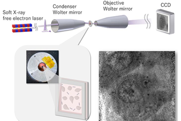
The microscope has two key components: a soft X-ray free electron laser; and highly precise Wolter mirrors, a type of mirror widely used in X-ray telescopes for observing space. The mirrors were made using technology created by lead author Satoru Egawa, assistant professor of the Research Center for Advanced Science and Technology at the University of Tokyo.

“A soft X-ray free electron laser provided pulse illumination at the speed of tens of femtoseconds (with one femtosecond being one-millionth of one-billionth of a second). The ultrashort duration of the radiation pulses enabled us to take an image before the structure of the living cell was altered by radiation damage,” explained Egawa. “We used Wolter mirrors for illumination and imaging. These mirrors provide a wide field of view, can withstand irradiation from the powerful lasers and exhibit no color distortion, making them ideal for observing samples at various wavelengths.”
Although soft X-ray free electron lasers have previously been used to study smaller viruses and bacteria, mammalian cells were too big to be studied this way. However, by using Wolter mirrors, the team could achieve a wider field of view and use a thicker sample holder which could hold larger cells. The resulting images showed details about carbon content in the cells that had not been seen through other methods, such as electron microscopy and fluorescence microscopy.
“It was surprising for us to find a carbon pathway between the nucleolus (a structure in the cell’s nucleus, involved in cell function and survival) and the nuclear membrane (which envelops the nucleus), which had not been observed with visible light microscopes,” said Egawa.
Brighter soft X-ray free electron lasers are available which would enable even clearer images with less grainy “noise.” By adding brighter lasers and more precise Wolter mirrors, the team hopes to upgrade the microscope so that it can observe more biochemical elements. With this it could also help to illuminate some of the vital reactions and interactions which take place within living cells.