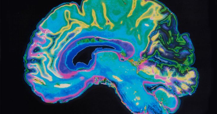
By combining noninvasive imaging techniques, investigators have created a comprehensive cellular atlas of a region of the human brain known as Broca’s area — an area critical for producing language.
The new technology will provide insights into the presence and spread of pathologic changes that occur in neurodegenerative illnesses — such as epilepsy, autism, and Alzheimer’s disease — as well as psychiatric illnesses.
Until now, scientific advances have not produced undistorted 3D images of cellular architecture that are needed to build accurate and detailed models. In new research published in Science Advances, a team led by investigators at Harvard-affiliated Massachusetts General Hospital, has overcome this challenge with detailed resolution to study brain function and health.
Using sophisticated imaging techniques — including magnetic resonance imaging, optical coherence tomography, and light-sheet fluorescence microscopy — the researchers were able to prevail over the limitations associated with any single method to create a high-resolution cell census atlas of a specific region of the human cerebral cortex, or the outer layer of the brain’s surface. The team created such an atlas for a human postmortem specimen and integrated it within a whole-brain reference atlas.


