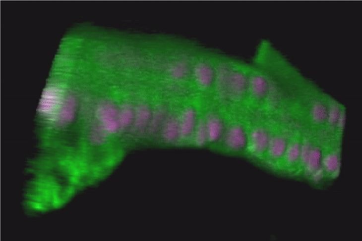
 The Parthenon-like structure. Most of the volume of epithelial cell bodies containing nuclei are aligned as a monolayer on the apical (upper) side. They extend pillarlike cell processes in a basal (downward) direction. At the basal (lower) side, the cytoplasm of epithelial cells spread planarly, creating a thin layer. Macrophage-like cells are found in the cavities between pillars. ©2024 Tetsuya Kojima, Graduate School of Frontier Sciences, The University of Tokyo
A little leg may reveal something big about how closely related insect species can drastically differ in body shape, according to a new study led by researchers at the University of Tokyo. The team used time-lapse microscopy to image the live cells of fruit flies and found a new structure, which forms and disappears in the final stages of a common fruit fly’s development, that appears to help guide a section of the fly’s leg into its final shape. The findings could lead to better understanding the mechanisms determining an insect body’s shape, as well as providing insight into processes shaping the bodies of other organisms.
In the current study, the research team set out to clarify how certain cells determine an organism’s final shape, using fruit fly leg development as a model. By imaging the live cells of the common fruit fly Drosophila melanogaster over several days to analyze changes in the cells during the last stages of development, the researchers found a transient structure in the fly’s leg involved in determining the limb’s final shape. They named this formation the “Parthenon-like structure” because of a resemblance to the ancient Greek ruin.
 Final shape formation of the leg through transient formation of the Parthenon-like structure. Scientists revealed that the final shape formation of the fruit fly leg proceeds through the transient formation of the Parthenon-like structure. ©2024 Tetsuya Kojima, Graduate School of Frontier Sciences, The University of Tokyo
“Cells change their shape more dramatically than ever thought during the final shape formation process,” said co-author Tetsuya Kojima, associate professor in the Department of Integrated Biosciences at the University of Tokyo’s Graduate School of Frontier Sciences. “Especially in the case of the fruit fly’s leg, cells transiently form the fascinating structure. Because the Parthenon-like structure is seen in tissues other than the leg and seems to appear in other insects, its transient formation may be a fundamental process that forms the final shape of the insect’s body.”
According to the researchers, the basic mechanisms of cell fate determination — how cells figure out which genes to express and contribute to the organism’s developed form — are conserved among closely related species. However, how these cells contribute to the form’s shape has remained elusive.
“Since shapes can differ dramatically between closely related species that are expected to share the basic mechanisms of cell fate determination, differences in final shape formation processes should greatly contribute to making the shape differences,” said co-author Reiko Tajiri, a researcher at the University of Tokyo at the time of the study and currently associate professor at Chiba University in Japan. “Understanding the mechanisms of final shape formation is of great importance to understanding the mechanisms of formation and diversification of organisms’ shapes.”
To understand the mechanisms of final shape formation, the researchers used an inverted confocal microscope — which images the specimens from underneath and allows for clearer visuals — to image the developing legs of fruit flies over several days. They specifically focused on the tarsus, or the segment of leg farthest from an insect’s body. The tarsus can appear flat and wide in male diving beetles or long and slender in mosquitoes. The tarsus also contains its own segments, which differ significantly across species.
 Overview of the final leg shape formation. The final leg shape formation process can be divided into five stages. (APF is an abbreviation for: after puparium formation.) ©2024 Tetsuya Kojima, Graduate School of Frontier Sciences, The University of Tokyo
“This diversity in morphology (form and structure) makes the insect tarsus a good model for studying the mechanisms of final shape formation and its diversification,” Kojima said. “In this study, we found unexpected and dramatic shape changes in epithelial cells — cells that line the surface of structures — and the basement membrane, which give rise to the structures that differentiate into the tarsal segments. We also saw interesting behavior of macrophage-like cells, which typically help clean up waste cells.”
Capturing nearly the whole shaping process of the adult tarsus during the fly’s pupal stage, the researchers observed that the epithelial cells changed from a column shape to be more cubelike. They said the cells further changed to “dramatically” form the unexpected, transient Parthenon-like structure. As that structure forms and then disappears, the tarsus’s diameter reduces rapidly. Following the reduction in diameter, the layer of epithelial cells thinned out.
“Our results contribute to elucidating the mechanism for the final shape formation of the adult tarsus,” Kojima said, noting that the team is now studying the formation and disappearance of the Parthenon-like structure in more detail to better understand how it contributes to making the final leg shape.
 Live imaging from Stage III to Stage IV. Cells dynamically change their shape and diameter, rapidly decreasing during Stage III. Cells rapidly going around are macrophage-like cells. (View the longer version of the video from Stage III to Stage V.) ©2024 Tetsuya Kojima, Graduate School of Frontier Sciences, The University of Tokyo